Danijela Županić [1] Jelena Vlašić-Tanasković [1] Ružica Šmalcelj [2] Petar Kes [2] Vesna Kušec [*] [3]

Show more about author

Introduction

 
Metabolic bone disorder occurs in most patients with chronic kidney failure treated by chronic dialysis primarily due to impaired calcitriol synthesis and increased secretion of parathyroid hormone (PTH) (1,2,3). Other factors, i.e. bone regulatory proteins, have also been implicated, e.g. bone morphogenetic proteins and osteoprotegerin (4). The course of the disease and treatment has an effect on the type of bone turnover disorder, resulting in either high or low bone remodeling (5). High bone turnover is characterized by increased and accelerated bone formation, and high bone resorption rate (6,7). The consequences on the skeletal system are greater osteoid volume, formation of woven bone, increased and intratrabecular resorption leading to osteopenia or osteoporosis with greater fracture risk (8,9). Osteomalacia or adynamic bone are characterised by low bone turnover, i.e. arrested renewal of bone tissue and microfracture repair also resulting in increased fracture risk (9). Duration of kidney failure and dialysis treatment, PTH secretion and its therapeutic control are important determinants of this bone disorder.
After successful kidney transplantation most metabolic disturbances are resolved, but normalization of parathyroid hypersecretion and disorder of bone metabolism may take 2-4 years (2,10,11,12). Increased bone turnover in kidney transplant recipients is related to previous dialysis duration, PTH levels and thetime since transplantation. The main condition is additionally aggravated by immunosuppressives and corticosteroids, the drugs deleterious to skeletal integrity which are administered in the post-transplant period (e.g. 13,14,15,16).
Assessment of the skeletal status and bone disorder in patients on chronic dialysis and kidney transplant recipients is accomplished by measurement of bone mineral density, histologic/histomorphometric analysis of bone biopsy and determination of biochemical bone markers (17). Products of bone cell activities are measured in serum and urine for indicators of bone formation and bone resorption. Investigation of bone markers has mostly shown increased bone turnover in patients on dialysis and also in kidney transplant patients (7,18,19,20). Comparison of bone markers and bone histology indicated that thebone disorder characterized by high bone turnover could be established by bone markers (21,22,23,24,25). In the case of low bone turnover, measurement of normal or decreased levels of bone markers was not found acceptable for diagnosis of this condition. The reports on clinical utility of bone markers in patients on chronic dialysis treatment and kidney transplant recipients differ depending on bone markers measured and patient population characteristics. In order to contribute to the better understanding of metabolicbone disorder, the aim of this studywas analysis of bone formation and bone resorption markers in two separate groups of patients, those on chronic hemodialysis treatment and after kidney transplantation with regard to specific risk factors of this disorder.
 

Materials and methods

 
Blood samples were collected in the course of routine monitoring of bone disorder in 79 patients (50 men, 29 women) on chronic hemodialysis and 36 patients (20 men, 16 women) with stable kidney transplant. Bone biopsy was not performed in these patients, but clinical data did not indicate adynamic bone. None of the patients had diabetes. Blood was withdrawn after an overnight fast and before hemodialysis treatment in patients with kidney failure, thesera separated and stored frozen (-20 oC) till assayed within one month of storage. The sera remaining after routine biochemical tests for patient monitoring were used in this investigation.
Hemodialysis patients underwent renal replacement treatment for4 hours 3 times a week, calcium concentration in dialysis fluid was on average 1.50 mmol/L (range 1.25-1.75 mmol/L). Standard therapy for these patients included phosphate binders (calcium carbonate, Sevelamer hydrochloride) and/or calcitriol according to clinical requirements and laboratory tests.
Immunosuppressive therapy in kidney transplant recipients consisted of Cyclosporin A, prednisone and azatioprine mofetil mycophenolate. The mean serum creatinine concentration for this patient group was 119.7 ± 20.5 μmol/L (range 79-168 μmol/L) and creatinine clearance was 70.2 ±13.5 mL/minute (range 51-112 mL/minute).
Data on age, duration of hemodialysis and post-transplant period for the patients comprised in this study are presented in Table 1. No difference existed between sexes regarding age, hemodialysis duration or post-transplant period for each group of patients.
 
Table 1. Clinical characteristics of the studied patient groups as presented by descriptive statistics (arithmetic mean = X, standard deviation = SD, range).
 
162_Zupanic_D._i_sur_Tablica_1
 
The following biochemical parameters were measured by commercial kits according to manufacturers’ recommendations: intact PTH (iPTH, IBL, Germany, reference range 1-6 pmol/L; for dialysis patients approximately 3x upper reference limit is recommended), bone alkaline phosphatase (Ostase BAP, IDS, Tyne&Wear, UK, reference limit <22 IU/L) as a bone formation marker, and C-terminal telopeptide of collagen type I (Crosslaps Serum, Nordic Bioscience Diagnostics, Herlev, Denmark, reference limit <0.9 ug/L) as abone resorption marker. In case of different reference ranges for sexes, the values for postmenopausal women were used as the highest result in healthy individuals.
Statistical analyses were performed by a standard statistical package (Microsoft® Office Excell) including descriptive statistics, correlation and regression assessment, student’s t-test (two-tail) for difference between means with significance level p<0.05.
 

Results

 
Data on PTH and bone markers showed that no difference existed as tested by t-test between sexes in each of the patient groups. Further statistical analyses were thus carried out for the entire group of patients on hemodialysis or kidney transplant recipients.
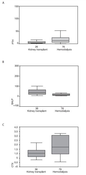
Based on the two tail test, patients on chronic hemodialysis were significantly older (p=0.0005) and were significantly longer treated by hemodialysis (p=0.04) as compared to kidney transplant recipients. Differencesin PTH and bone markerlevels were tested by t-test between patient groups (Table 2). Patients on chronic hemodialysis had significantly lower BALP, higher crosslaps and PTH in comparison to kidney transplant recipients (Figure 1). Graphic displays also enable observation of greater ranges for PTH and crosslaps in patients on hemodialysis, while variations are less pronounced in kidney transplant recipients. In a proportion of patients in both patients groups, PTH and bone markers were above the reference range, and percentages of increased results were indicated for each parameter and each patient group in Table 2. For PTH and BALP higher percentages of increased results were found in kidney transplant recipients and for crosslaps in chronic hemodialysis patients. When evaluating PTH results, it should be taken into account that reference limits are different, i.e. approximately 3x higher for chronic dialysis patients.
 
Table 2. Results of t-test between patients on chronic hemodialysis and kidney transplant recipients. Data are presented as mean and standard deviation, patient population size (n) and percentage of increased values in brackets. Please note that increased PTH for patients on chronic hemodialysis are greater than 18 pmol/L and in kidney transplant recipients greater than 6 pmol/L.
 
162_Zupanic_D._i_sur_Tablica_2
 
 
bm-16-137-f1
Figure 1. Data on PTH, BALP and crosslaps were significantly different between kidney transplant recipients (kidney transplant) and patients on chronic dialysis treatment (hemodialysis) as presented by box-and-whiskers plots.
 
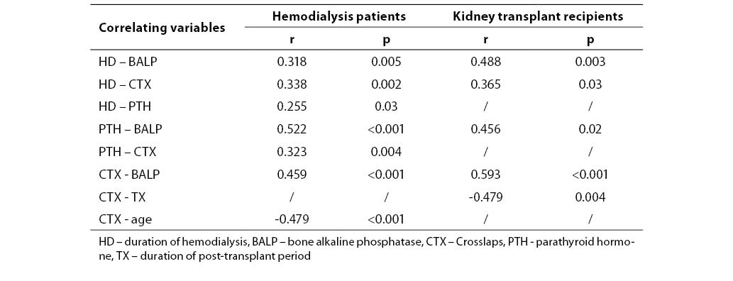
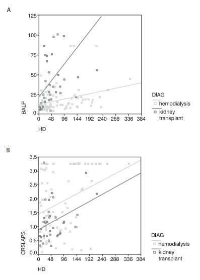
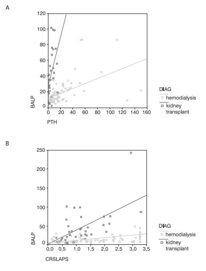
Correlations were assessed in order to examine relationships between biochemical parameters and investigated clinical data. In patients on chronic hemodialysis, the duration of hemodialysis treatment correlated significantly and positively with PTH, BALP and crosslaps. Age was found to correlate negatively with crosslaps. Statistically significant and positive correlations existed for PTH with BALP and crosslaps, and BALP with crosslaps. In kidney transplant recipients previous hemodialysis duration correlated significantly and positively with BALP and crosslaps, post-transplant period negatively with crosslaps. Biochemical parameters were also significantly and positively correlated, PTH with BALP, and BALP with crosslaps. Only statistically significant correlations and linear regression equations for pairs significant in both patients groups are presented in Table 3. Figures 2 and 3 show differences in associations of analyzed parameters between the two patient groups. Relationship of BALP with hemodialysis duration demonstrated lesser slope in patients on chronic hemodialysis and a steep increase of BALP with previous hemodialysis duration in kidney transplant recipients. Higher BALP levels in kidney transplant recipients were already reported in Figure 1 and Table 2. Increase of crosslaps in relationship to hemodialysis duration showed a parallel pattern, but higher values in patients on chronic hemodialysis (Figure 2). In relationships of PTH with BALP and crosslaps with BALP, different patterns of associations between patients on chronic hemodialysis and kidney transplant recipients were observed, with steeper slopes for kidney transplant recipients (Figure 3).
 
Table 3. Statistically significant correlations for pairs of parameters in patients on chronic hemodialysis and kidney transplant recipients presented by correlation coefficient (r) and level of significance (p). Linear regression equations are included only for pairs of variables found to correlate significantly for both patient groups.
 
162_Zupanic_D._i_sur_Tablica_3
 
bm-16-137-f2
Figure 2. Plots of BALP and crosslaps (CRSLAPS) in relationship to duration of dialysis treatment (HD) for patients on chronic dialysis (hemodialysis) and kidney transplant recipients (kidney transplant) with linear regression indicate different associations of analyzed parameters for patient groups.
 
bm-16-137-f3
Figure 3. Plots of BALP with PTH and BALP with crosslaps (CRSLAPS) for patients on chronic dialysis (hemodialysis) and kidney transplant recipients (kidney transplant) with linear regression indicate different associations of analyzed parameters for patient groups.
 

Discussion

 
In this investigation bone metabolism was compared in patients on chronic hemodialysis treatment and kidney transplant recipients as assessed by PTH and bone markers, and also with regard to age, sex, hemodialysis duration and post-transplant period. Bone turnover was increased in both patient groups, in particular bone resorption. Duration of hemodialysis and PTH were recognized as determinants of increased bone turnover in both groups, and decrease inbone resorption was related to post-transplantation period. In this bone disorder, sex and age were not established as risk factors for impaired bone turnover.
The measured biochemical parameters PTH and crosslaps were significantly higher in patients on chronic hemodialysis in comparison to kidney transplant recipients, and accordingly the percentages of increased crosslaps were higher. The proportion of increased PTH results was lowerin chronic hemodialysis patients, which can be explained by higher reference limit than in subjects with normal renal function (i.e. approximately >3x normal). The fact that increased PTH was found in only 35% of chronic hemodialysis patients also suggested efficient clinical control of PTH secretion. Increased bone turnover in patients on chronic hemodialysis can be explained by the metabolic bone disorder that developed already in the course of deterioration of kidney function (26). The main determinant of accelerated bone turnover is PTH hypersecretion, while increased bone markers are a consequence of both increased, bone cell activity and accumulation of their products due to impaired kidney clearance. An unexpected finding in this study was higher, and in greater percentage increased, bone alkaline phosphatase in kidney transplant recipients than in patients on chronic hemodialysis. This was in contrast with PTH and crosslaps results for the two patient groups. Alkaline phosphatase is not cleared by the kidney and thus not increased in subjects with impaired kidney function. Measurement of bone alkaline phosphatase is thus considered an appropriate marker for assessment of bone metabolism in patients on chronic dialysis (7,27,28). Possible cause of higher BALP in kidney transplant recipients could be the effect of immunosuppressive drugs (cyclosporine A) administered after kidney transplantation which may induce a rise in alkaline phosphatase (29,30). In the early post-transplant period a transient stimulation of bone remodeling was associated to cyclosporine A and decreasing prednisone doses (31). The observed higher BALP values in kidney transplant recipients could be attributed to immunosuppresive therapy but might also be a specific trait of bone metabolism disorder in this patient population. It has beeniswell established that renal osteodystrophy and bone disorder in patients on chronic dialysis and with kidney transplant arecharacterized by great variability in clinical forms, giving rise to a diagnostic and therapeutic problem (17).
Positive association of bone formation and resorption markers in both patient populations indicated the coupling of the bone cell processes in metabolic bone disorder (7,32,33). Differences between the two patient groups in relationships of hemodialysis duration, PTH and crosslaps with BALP were strongly influenced by higher BALP in kidney transplant recipients resulting in greater slope in associations as compared to thedata for patients on hemodialysis.
The correlations found between biochemical parameters and other patient data indicated similarities and differences in characteristics of chronic hemodialysis treatment and post-transplant disorder. Duration of hemodialysis and PTH secretion are determinants of increased bone turnover in patients on chronic hemodialysis treatment and also in kidney transplant recipients (6,20,34,35), as observed insignificant positive correlations in this study. In kidney transplant recipients, previous treatment by hemodialysis correlatedwith the investigated bone markers, but not with PTH. This could be due to moderately increased PTH levels in this patient group, stressing the importance of good control of parathyroid function in the course of the disease (36). In turn, PTH correlated only with BALP, and not with thebone resorption marker crosslaps. Previous hemodialysis duration is a risk factor for increased bone turnover after kidney transplantation, i.e. the pre-transplant bone disorder continues after kidney transplantation (13,14,37). Correlations of hemodialysis duration with crosslaps for both patient groups demonstrated that duration of kidney failure wascorrespondingly detrimental for increased bone resorption (6), which was more pronounced in chronic hemodialysis patients than in kidney transplant recipients. As previously stated, this was primarily due to higher PTH levels found in patients treated by chronic hemodialysis. Similar to patients treated by chronic hemodialysis, PTH level is also an important determinant of increased bone turnover after kidney transplantation (2,13,14). This was not confirmed in this study and was probably a consequence of thespecific patient group and its size. After successful kidney transplantation, bone turnover was normalized in the post-transplant period (13), as observed innegative correlation with bone resorption marker in this investigation and also reported in our previous studies (37,38).
The fact that chronic hemodialysis patients were significantly older and were treated by hemodialysis for longer period than the kidney transplant patient population should not be disregarded. This mostly reflects that patients not fulfilling criteria for transplantation were older and thus longer treated by chronic hemodialysis, which are synergistic factors for disorder of bone metabolism.
Analysis of biochemical parameters in the two patient groups showed no difference between sexes. Bone markers in normal population differ between sexes both in adults and children. The negative influence of female sex on skeletal integrity is also observed in the postmenopause and is recognized as risk factor for osteoporosis. Difference between sexes in patients on chronic hemodialysis or kidney transplant recipients was not found by all investigators (39), thus implying aninvolvement of other risk factors of bone disease as opposed to generally accepted risk factors for osteoporosis.
Most of the relationships between bone markers, PTH and hemodialysis duration are common and similar to patients on chronic hemodialysis and kidney transplant recipients (10). This was reflected in correlation coefficients, which were of identical direction and similar magnitude in both patient groups.
A particular finding for patients on chronic hemodialysis was the negative correlation of age and the bone resorption marker crosslaps. Older age is a risk factor for osteoporosis, although not generally confirmed to be of significance in patients on chronic hemodialysis. In this study, bone resorption was found to be less pronounced in older patients. Similar association was not found for PTH levels.
Measurement of bone markers together with PTH can be considered a useful clinical tool in assessment and follow-up of metabolic bone disorders related tochronic hemodialysis treatment and kidney transplantation (40). It also enables insight into risk factors of this bone disorder and investigation into variations of itsclinical presentations.

Notes

This manuscript has been submitted in English. Croatian translation is the responsibility of the Editorial.

References

1.     Hruska KA, Teitelbaum SL. Renal osteodystrophy. N Engl J Med 1995;333:166-73.
2.     Parfitt AM. The hyperparathyroidism of chronic renal failure: a disorder of growth. Kidney Int 1997;52:3-9.
3.     Parfitt AM. A structural approach to renal bone disease. J Bone Miner Res 1998;13:1213-20.
4.     Hruska KA, Saab G, Chaudhary LR, Quinn CO, Lund RJ, Surendran K. Kidney-bone, bone-kidney, and cell-cell communication in renal osteodystrophy. Semin Nephrol 2004;24:25-38.
5.     Ferreira A. Development of renal bone disease. Eur J Clin Invest 2006;36(Suppl 2):2-12.
6.     Avbersek-Luznik I, Balon BP, Rus I, Marc J. Increased bone resorption in HD patients: is it caused by elevated RANKL synthesis? Nephrol Dial Transplant 2005;20:566-70.
7.     Cavalier E, Delanaye P, Collette J, Krzesinski JM, Chapelle JP. Evaluation of different bone markers in hemodialyzed patients. Clinica Chimica Acta 2006, in press
8.     Piraino B, Chen T, Cooperstein L, Segre G, Pushett J. Fractures and vertebral bone mineral density in patients with renal osteodystrophy. Clin Nephrol 1988;30:57-62.
9.     Cunningham J, Sprague SM, on behalf of the Osteoporosis Work Group. Osteoporosis in chronic kidney disease. Am J Kidney Dis 2004;43:566-71.
10.   Kanis JA, Cundy TF, Hamdy NA. Renal osteodystrophy. Baillieres Clin Endocrinol Metab 1988; 2: 193-241.
11.   Carlini RG, Rojas E, Weisinger JR, Lopez M, Martinis R, Arminio A, et al. Bone disease in patients with long-term renal transplantation and normal renal function. Am J Kidney Dis 2000; 36: 160-6.
12.   Kodras K, Haas M. Effect of kidney transplantation on bone. Eur J Clin Invest 2006;36(Suppl 2):63-75.
13.   Massari PU. Disorders of bone and mineral metabolism after renal transplantation. Kidney Int 1997;52:1412-21.
14.   Brandenburg VM, Westenfeld R, Ketteler M. The fate of bone after renal transplantation. J Nephrol 2004;17:190-204.
15.   Cruz DN, Wysolmerski JJ, Brickel HM, Gundberg CG, Simpson CA, Mitnick MA, et al. Parameters of high bone-turnover predict bone loss in renal transplant patients: a longitudinal study. Transplantation 2001;72:83-8.
16.   Maalouf NM, Shane E. Osteoporosis after solic organ transplantation. J Clin Endocrinol Metab 2005;90:2456-65.
17.   Martin KJ, Olgaard K on behalf of the Bone Turnover Work Group. Diagnosis, assessment, and treatment of bone turnover abnormalities in renal osteodystrophy. Am J Kidney Dis 2004;43:558-65.
18.   Nakanishi M, Yoh K, Uchida K, Maruo S, Rai SK, Matsuoka A. Clinical usefulness of serum tartarate-resistant fluoride-sensitive acid phosphatase activity in evaluating bone turnover. J Bone Min Metabolism. 1999; 17: 125-30.
19.   Ziolkowska H, Paniczyk-Tomaszewska M, Debinski A, Polowiec Z, Sawicki A, Sieniawska M. Bone biopsy results and serum bone turnover parameters in uremic children. Acta Paediatrica 2000; 89: 666-71.
20.   Šmalcelj R, Kušec V, Slaviček J, Barišić I, Glavaš-Boras S. Biochemical markers of bone metabolism in patients on chronic dialysis. Period Biol 2000;102: 55-8.
21.   Urena P, Ferreira A, Kung VT, Moreeux C, Simon P, Ang KS, et al. Serum pyridinoline as a specific marker of collagen breakdown and bone metabolism in HD patients. J Bone Miner Res 1995;10:932-9.
22.   Mazzaferro S, Pasquali M, Ballanti P, Bonucci E, Costantini S, Chicca S, et al. Diagnostic value of serum peptides of collagen synthesis and degradation in dialysis renal osteodystrophy. Nephrol Dial Transplant 1995;10:52-8.
23.   Coen G, Ballanti P, Bonucci E, Calabria S, Centorrino M, Fassino V, et al. Bone markers in the diagnosis of low turnover osteodystrophy in haemodialysis patients. Nephrol Dial Transplant 1998;13:2294-302.
24.   Kušec V, Potočki K, Šmalcelj R, Puretić Z, Kes P, Gašparov S, et al. Praćenje poremećaja metabolizma kosti nakon transplantacije bubrega. Documenta urologica 2001-2002; 1: 3-7.
25.   Kušec V, Šmalcelj R. Značenje biokemijskih pokazatelja koštane pregradnje u bolesnika na kroničnoj dijalizi i nakon transplantacije bubrega. Acta Med Croatica. 2004;58(1):51-7.
26.   Hutchison AJ, Whitehouse RW, Boulton HE, Adams JE, Mawer EB, Freemont TJ, et al. Correlation of bone histology with parathyroid hormone, vitamin D3 and radiology in end-stage renal disease. Kidney Int 1993;44:1071-7.
27.   Inaba M, Nagasue K, Okuno S, Ueda M, Kumeda Y, Imanishi Y, et al. Impaired secretion of parathyroid hormone, but not refractorinessof osteoblast, is a major mechanism of low bone turnover in hemodialyzed patients with diabetes mellitus. Am J Kidney Dis 2002; 39:1261-9.
28.   Ueda M, Inaba M, Okuno S, Maeno Y, Ishimura E, Yamakava T, et al. Serum BAP as the clinically useful marker for predicting BMD reduction in diabetic hemodialysis patients with low PTH. Life Sci 2005;77:1130-9.
29.   Wilmink JM, Bras J, Surachno S, v Heyst JLAM, v d Horst JM. Bone repair in cyclosporine treated renal transplant patients. Transplant Proc 1989; 21:1492-94.
30.   Briner VA, Landmann J, Brunner FP, Thiel G. Cyclosporine A-induced transient rise in plasma alkaline phosphatase in kidney transplant patients. Transplant Int 1993; 6:99-107.
31.   Westeel FP, Mazuouz H,Ezaitouni F, Hottelart C, Ivan C, Fardellone P, et al. Cyclosporine bone remodeling effectprevents steroid osteopenia after kidney transplantation. Kidney Int 2000;58:1788-96.
32.   Malyszko J, Wolczynski S, Malyszko JS, Konstantynowicz J, Kaczmarski M, Mysliwiec M. Correlations of new markers of bone formation and resorption in kidney transplant recipients. Transplantation Proceedings 2003;35:1351-54.
33.   Maeno Y, Inaba M, Okuno S, Yamakawa T, Ishimura E, Nishitzawa Y. Serum concentration of cross-linked N-telopeptides of type I collagen: New marker for bone resorption in hemodialysis patients. Clin Chem 2005;51:2312-7.
34.   Cueto-Manzano AM, Konel S, Hutchinson AJ, Crowley V, France MW, Freemont AJ, et al. Bone loss in long-term renal transplantation: histopathology and densitometry analysis. Kidney Int 1999;55:2021-9.
35.   Wittersheim E, Mesquita M, Demulder A, Guns M, Louis O, Melot C, Dratwa M, Bergmann P. OPG, RANK-L, bone metabolism, and BMD in patientson peritoneal dialysis and hemodialysis. Clin Biochem 2006;39:617-22.
36.   Gomes CP, Barreto Silva, Leite Duarte ME, Dorigo D, da Silva Lemos CC, Bregman R. Bone disease in patients with chronic kidney disease under conservative management. Sao Paulo Med J 2005;123:83-7.
37.   Kušec V, Šmalcelj R, Cvijetić C, Rožman R, ŠkrebF. Determinants of reduced bone mineral density and increased bone turnover after kidney transplantation. Croatian Med J 2000;41(4):396-400.
38.   Kusec V, Smalcelj R, Puretic Z, Szekeres T Interleukin-6, transforming growth factor-beta 1, and bone markers after kidney transplantation. Calcif Tissue Int. 2004;75:1-6.
39.   Julian BA, Laskow DA, Dubowsky J, Dubowsky EV, Curtis JJ, Quarles LD. Rapid loss of vertebral mineral density after renal transplantation. N Engl J Med 1991;3225:544-50.
40.   Schwarz C, Suzzbacher I, Oberbauer R. Diagnosis of renal osteodystrophy. Eur J Clin Invest 2006;36(Suppl 2):13-22.