The burden of heart failure
Heart failure (HF) represents the inability of the heart to respond to the circulatory demand of the organism (1). The main causes of HF are hypertension, ischaemic and valvular injuries whereas toxic, metabolic or genetic origins are less common. In addition to the initial abnormality, secondary changes occur over the course leading to multi-organ impairment (2). The prevalence of HF is important and more than 20 million people worldwide are estimated to suffer from HF (3). HF is increasing because of an ageing population, the success in prolonging survival in patients suffering of coronary events, and the success in postponing coronary events by effective prevention in those at high risk or those who have already survived to a first event (4). HF appears also as a deadly and costly disorder, carrying an overall worse prognosis (5). Thus, HF represents a major public health problem with a high degree of morbidity (6).In addition, the economical impact of HF is tremendously important for healthcare systems (7). In United Kingdom, Stewart et al. have estimated that the annual direct cost of HF in year 2000 was about 1.9% of the total expenditure of the National Health Service (7).
Cardiac hypertrophy and remodeling
Alteration in the heart workload leads to major changes in myocardial structure and function. Increased workload induces adaptive mechanisms maintaining adequate blood flow through the pulmonary and systemic circulations. Ultimately, prolonged cardiovascular stress leads to myocardial deterioration and heart failure. Cardiac remodeling is characterized by cardiac hypertrophy, fibrosis and inflammatory process. Exercise conditioning, which chronically increases the heart work, leads to some degree of myocardial hypertrophy. This physiologic hypertrophy is characterized by increases in both length and width of cardiomyocytes, and is referred to “proportional” (8). Pathological myocardial hypertrophy is induced mainly by hypertension, valvular dysfunction and loss of myocytes following ischemic damage (9). Some genetic alterations may also lead to pathological hypertrophy (10). At the early stages of chronic pressure overload, the cardiomyocytes hypertrophy is characterized by an increase in cell size and stimulation of protein synthesis. The assembly of sarcomeres occurs in parallel, resulting in widening of the cardiomyocytes. Typically in chronic arterial hypertension this leads to reduced left ventricular volume and increased wall thickness, referred to a concentric hypertrophy (11). The hypertrophy related to the early stages of cardiomyocytes hypertrophy is also characterized by the reactivation of the fetal gene program, with reactivation of b myosin heavy chain, α-actin and ANP gene expression (12). The expression of other genes already expressed in the normal adult myocardium, such as BNP and endothelin genes, are also increased. In more advanced chronic stages, the cardiac chambers dilate together with a thinning of the ventricular wall. The sarcomeres are then organized in series causing cell elongation; this long standing process leads to dilation of the heart cavities, characteristics of the eccentric dilation of HF.Heart failure is also characterized by a loss of myocytes due to cell apoptosis as well as progressive fibrotic processes. The occurrence of fibrosis is related to the proliferation of cardiac fibroblasts, with simultaneous collagen deposition and will contribute to the loss of cardiac function (13-15). Cardiac remodeling will also affect electrophysiological properties and contribute to cardiac arrhythmia.
Heart failure, neurohormonal activation and biomarkers
Most of the biomarkers with potential diagnosis and prognosis applications derive from the neurohormonal response to the failing myocardium. Indeed, neurohormonal activation plays a significant role in myocardial and multi-organ adaptations to HF (16,17). The use of biomarkers for the diagnosis of patients suspected of HF is constantly raising and is now part of daily practices in industrialized countries (6,18). Thus, rapid and sensitive tests for B-type natriuretic peptide (BNP) and the biologically inactive N-terminal fragment (NT-proBNP) are routinely used and are included in the ESC guidelines as the next step beyond clinical examination, ECG, chest-X-ray and echocardiography for the classification of patients suspected to present HF. Biomarkers may also fulfil complementary information for the evaluation of the disease severity, the prognosis estimation and for treatment selection and biomarkers of inflammation and cardiac remodeling may provide additional information to natriuretic peptides testing (19). As previously mentioned, the remodeling and fibrosis of the heart plays an important role in the progression of heart failure. Biomarkers related to cardiac hypertrophy, cardiac fibrosis and remodeling of the extracellular matrix may provide valuable information for the risk stratification of HF patients. Inflammation is stimulating cardiac remodeling and fibrosis and thus participates in the pathogenesis and progression of HF. Therefore, biomarkers related to the inflammatory response in HF are intensively evaluated (20). Several cytokines and biomarkers related to the activation of neutrophils and macrophages have been investigated for their ability to contribute to the risk stratification of HF patients.
Biomarkers of inflammation and cardiac remodeling already available for testing
Cytokines
The circulating levels of cytokines are enhanced in the failing myocardium (21). Thus, after following the initial heart insult, the increased production of proinflammatory cytokines may challenge the surrounding tissue through propagation of the inflammatory response and direct effects on the cardiac myocyte structure and function (22). Pro-inflammatory cytokines such as TNF-α, IL-6, IL-1, and IL-18 appear to cause cardiomyocytes apoptosis and necrosis as well as cells hypertrophy. The increase of circulating cytokines stimulated by HF may also predict long-term outcomes (20,22). Testing for cytokines remains challenging and difficult to translate in routine laboratories as the assays commonly used are based on enzyme linked immunosorbent assay (ELISA) format and remain expensive.
C-reactive protein
The levels of C-reactive protein (CRP) are enhanced by HF (23). Higher C-reactive protein predicts worse prognosis in acute heart failure only in non infected patients (24). Recent studies have reported a potential added-value of CRP for the risk prediction of adverse clinical events independently of natriuretic peptides (25). The potential advantage of CRP testing is its availability within the majority of laboratories through automated assays.
Myeloperoxidase
Myeloperoxidase (MPO) is a biomarker of inflammation and oxidative stress produced by neutrophils, monocytes, and endothelial cells. The levels of MPO are stimulated by HF and may appear as an independent predictor of mortality in HF. In a study including 667 patients presenting to the emergency department with dyspnea and observed them for 1 year, Reichlin et al. have measured the circulating levels of MPO (26). In their study, the increases in MPO significantly contributed, after adjustment for cardiovascular risk factors in multivariable Cox proportional hazard analysis, to the prediction of 1-year mortality. MPO testing was first presented as ELISAs but nowadays the tests menu of some automated platforms is offering access to this parameter.
Copeptin
Copeptin is the C-terminal fragment of the arginine vasopressin (AVP) (27). Copeptin levels are enhanced in response to stress states and infectious diseases. Recent studies have demonstrated the raise of Copeptin levels in case of HF and its potential as a marker for mortality and morbidity (28). Neuhold et al. showed in a cohort of more than 700 HF patients, that copeptin concentrations were related to NYHA functional class (29). In addition, their study highlighted copeptin as the most potent single predictor of mortality in patients with NYHA functional class II and class III. In the OPTIMAAL study, Voors et al. have compared copeptin with BNP and NT-proBNP for prediction of death or a composite cardiovascular endpoint in patients who developed HF after an acute myocardial infarction (28). In this study, the ROC curves indicated that copeptin measurement at baseline was a stronger predictor of mortality compared with both BNP and NT-proBNP (28). The copeptin assay can be available as ELISA but has also been developed on an automated platform. Therefore, the access to copeptin testing can be proposed by some reference laboratories.
Endothelin
Endothelin-1 (ET-1) is a 21 amino acids peptide and is one of the most potent vasoconstrictors (30). ET-1 is synthesized as an inactive 212 amino acid preprohormone, preproET-1. The latter is cleaved by endopeptidases to the 39–amino acid, big ET-1 (31). A final cleavage step, performed by endothelin converting enzyme-1, converts big ET-1 to the 21–amino acid product ET-1. Effects of ET-1 are mediated through stimulation of 2 subtypes of receptors, endothelin receptor subtype A and endothelin receptor subtype B. ET-1 is produced mostly in endothelial cells, kidney and central nervous system (31). Beside a transient nitric oxide mediated vasorelaxant effect, ET-1 is a potent vasoconstrictor. ET-1 exert also direct actions on the heart, such as chronotropic and inotropic effects, decrease of cardiac output, stimulation of myocardial hypertrophy ET-1 and induction of collagen synthesis response in cardiac fibroblasts (32). ET-1 levels are increased in HF patients and Van Beneden et al. have demonstrated in severe HF patients (33). Big ET-1 and ET-1 are strong independent predictors of survival in such and better for this purpose than natriuretic peptides or their pro-peptides (33). Therefore, these markers allow easily identifying a population with high risk mortality eligible for more aggressive therapies. As for cytokines, testing for endothelins remains challenging and difficult to translate in routine laboratories as the assays are based on ELISA format and remain expensive. Thus, only some reference laboratories are able to propose these biomarkers.
Future potential biomarkers of cardiac remodeling
Neopterin
Neopterin belongs to the chemical group known as pteridines and is produced by macrophages upon stimulation with cytokines (34). Neopterin is indicative of a pro-inflammatory immune status (34). Neopterin concentrations have been evaluated in HF patients (35). The authors have examined a group composed of 47 patients with NYHA class II and III HF and 20 healthy volunteers. HF patients had higher basal concentrations of neopterin than the control group. In a 12-month observation, the authors have also reported a relationship between neopterin concentration and HF progression. In another study, Sasaki et al. have determined the serum neopterin concentration was measured in 198 patients with chronic HF and 62 control subjects (36). In their study, the concentration of neopterin was increased with advancing NYHA classes. Furthermore the high neopterin group had a significantly higher incidence of cardiac events than low neopterin group. Recently, in a larger cohort Nazer et al. have evaluated the prognostic ability of neopterin testing (37). Thus, they have assessed the relationship between neopterin and hospitalization for HF, and for death or HF over a mean follow-up of two years in 3946 subjects with acute coronary syndrome and demonstrated that neopterin levels are an independent predictor of HF hospitalization, and improve risk prediction over and above conventional biomarkers. Hennig et al. have also recently proposed a potential role for inflammatory biomarkers and neopterin for prediction of right ventricular failure after implantation of a left ventricular assist device. (38). Neopterin testing is mainly based on manual ELISA devoted to research use. Raising evidence may facilitate the translation of neopterin testing to more automated analyzers.
Galectin-3
Galectin-3, a member of the lectin family, contains a carbohydrate-recognition-binding domain (CRD) of about 130 amino acids that enable the specific binding of β-galactosides (39). Galectin-3 expressed in the nucleus, cytoplasm, mitochondrion, cell surface, and extracellular space. This protein has been shown to be involved in cell adhesion, cell activation, cell growth and differentiation, cell cycle, and apoptosis. Recent evidence suggests that galectin-3 is up-regulated in hypertrophied hearts and may represent an important mediator for the development of fibrosis and cardiac remodeling (39). Van Kimmenade et al. have determined the circulating concentrations of galectin-3 as well as NT-proBNP and apelin in 599 patients presenting with dyspnea at emergency department (40). The galectin-3 levels were significantly higher in subjects with HF compared with those without. Receiver operating characteristic analysis for mortality prediction showed that, for 60-day prognosis, galectin-3 had the greatest area under the curve in comparison to NT-proBNP, BNP and apelin. Shah and coworkers have also measured galectin-3 in patients presenting to ED with acute dyspnea and confirmed this prognosis ability of galectin-3 (41). Thus,in their study galectin-3 remained a significant predictor of 4-year mortality independent of echocardiographic markers of risk. The evidence related to galectin-3 testing is accumulating quickly which is leading to its transfer to several automated analyzers as well as point-of-care devices.
GDF-15
Growth differentiation factor 15 (GDF15), a protein belonging to the transforming growth factor beta superfamily, has a role in regulating inflammatory and apoptotic pathways in injured tissues and during disease processes. GDF15 acts anti-apoptotic and pro-hypertrophic in adult cardiomyocytes (42). The circulating concentration of GDF-15 was measured at baseline and at 12 months in patients randomized in the Valsartan Heart Failure Trial (43). Higher levels of GDF15 were associated with features of worse HF and biomarkers of neurohormonal activation, inflammation, myocyte injury, and renal dysfunction. Baseline GDF-15 levels were also associated with the risks of mortality and remained independently related to mortality in a Cox regression model that included clinical prognostic variables, B-type natriuretic peptide, high-sensitivity C-reactive protein, and high-sensitivity troponin T. In another study high values of GDF15 were also reported in HF patients with normal ejection fraction and may support diagnosis of HF (44). The GDF15 testing is mainly devoted to research use now but may enter the menu of some automated analyzers in a near future.
Osteopontin
Osteopontin (OPN) is is a macrophage-derived, RGD-containing glycoprotein with cytokine-like, chemotactic, and proadhesive properties. OPN is also an extracellular structural protein and an organic component of bone. The over-expression of OPN has been related to dilated cardiomyopathy (45). The myocardial expression of OPN has been described to increase with the severity of HF (46). The plasma levels of OPN have been related to HF, its worsening course and the efficiency of left ventricular assisted device (47). OPN assays are based on ELISA format and remain expensive. Thus, only some reference laboratories are able to propose these biomarkers.
Osteoprotegerin
Osteoprotegerin (OPG) is a cytokine receptor and a member of the tumor necrosis factor receptor superfamily. OPG and its ligand, the receptor activator of nuclear factor-κB ligand (RANKL), are regulating bone remodeling (48). In the Controlled Rosuvastatin Multinational Trial in HF (CORONA) population, randomly assigned to 10 mg rosuvastatin or placebo, OPG levels added independent predictive information for the worsening of HF (49). The plasma OPG levels were also assessed in 1,229 patients with HF recruited from 51 clinical centers and included in the Gruppo Italiano per lo Studio della Sopravvivenza nell’Infarto Miocardico-Heart Failure (GISSI-HF) trial (50). After adjustment for conventional risk markers, OPG remained a significant predictor of death. OPG assays are based on ELISA format and remain expensive. Thus, only some reference laboratories are able to propose these biomarkers.
ST-2
ST2 is an interleukin-1 receptor family member with transmembrane(ST2L) and soluble (sST2) isoforms (51). ST2 was originally described in the context of inflammatory and autoimmune diseases. However, after the identification of IL-33 as the functional ligand for ST2, and conceptualization of the role of ST2/IL-33 signaling in cardiac remodeling, sST2 has emerged as a potential novel cardiovascular biomarker for the presence of ventricular biomechanical overload (52). Rehman et al. have characterized ST2 in patients with acute HF (53). The ST2 values correlated with the severity of HF, the left ventricular ejection fraction, BNP and NT-proBNP (53). The ST2 levels at presentation were higher among patients who died by 1 year. In a multivariable Cox model containing established clinical and biochemical predictors, ST2 remained a predictor of mortality. When both ST2 and natriuretic peptides were elevated, the highest rates of death were observed in cumulative hazard analysis. Mueller et al. have also determined plasma concentrations of soluble ST2 in patients with DHF attending the ED (54). In multivariable Cox proportional-hazards regression analyses, an sST2 plasma concentration in the upper tertile at presentation was a strong and independent predictor of all-cause mortality after one year of follow-up. ST2 levels were also evaluated in the PRAISE-2 heart failure trial. In patients with severe chronic NYHA class III to IV HF, the change in ST2 levels is an independent predictor of subsequent mortality or transplantation (55). ST2 testing is mainly devoted to research use now but may enter the menu of some automated analyzers in a near future.
Urocortin
Urocortin (UCN) is a stress response peptide containing 40 amino acids belonging to the corticotrophin releasing factor (CRF) peptides family (56). In 2005, J. Burnett Jr. has presented UCN system as a component of the neurohormonal response to HF (57). UCN is able to induce hypertrophy of cardiac cells and immunohistochemical studies have demonstrated that UCN is expressed more abundantly in the diseased heart, dilated cardiomyopathy and hypertrophic cardiomyopathy, than in the normal heart (58,59). In contract, several studied have suggested that UCN treatment may have hemodynamic benefits in HF (57,60). Nevertheless, the data related to the circulating levels of UCN in HF are limited. Indeed, Ng and coworkers have reported that plasma levels of UCN were elevated in HF (61). They also shown that UCN levels increased with the NYHA class, especially in its early stage, were negatively correlated to Nt-proBNP (61). We have also demonstrated that the plasma concentrations of UCN are increased in HF patients in comparison to healthy individuals (62). However, our results shown that circulating concentrations of UCN are increased in HF patients, in both NYHA I-II and NYHA III-IV functional class subgroups and were contrasting with Ng et al. Wright et al. have recently reported that increased UCN levels in HF, an inverse relation with LVEF and a linear increase with NYHA (63). UCN assays are based on ELISA format and remain expensive. Thus, only some reference laboratories are able to propose these biomarkers.
Urotensin-II
Urotensin II (UII) is a cyclic undecapeptide, first isolated from teleost fish and later identified in mammals (64). UII possesses multiple effects on the cardiovascular system, mediated through activation of G-protein coupled receptors. In rat thoracic aorta, incubated in vitro, UII has been reported to be the most vasoconstrictor described so far. The expression of UII is increased in patients with end-stage HF in comparison to controls (65). The ability of UII to act also as an autocrine/paracrine factor participating to cardiac hypertrophy and fibrosis is also raising (64,66). Indeed, UII has hypertrophic effect on cardiomyocytes and may be implicated in myocardial fibrogenesis through increased collagen synthesis by cardiac fibroblasts (67). Thus, Richards et al. found that plasma UII was elevated in HF patients, but the study was restricted to NYHA class IV patients (68). In such a group, UII correlated significantly with ET-1 and adrenomedullin but not with NT-proBNP. Later, we have also demonstrated that the circulating levels of UII are increased in HF patients in comparison to healthy individuals (69). Moreover, we showed that plasma UII levels were related with the NYHA functional classes, the LVEF, as well as with established neurhormonal biomarkers of HF such as BNP and Big-ET-1. These results were in agreement with the work of Douglas and co-workers who have previously reported that the myocardial expression of UII correlated significantly with left ventricular end diastolic dimension and LVEF (65). As well as for ET-1, the early activation of UII may participate to myocardial hypertrophy to prevent the loss of systolic function but may also stimulate deleterious fibrosis and remodeling of the myocardium. UII assays are based on ELISA format and remain expensive. Thus, only some reference laboratories are able to propose these biomarkers.
Perspectives: several challenges before the translation to daily practices
Our article has presented some candidate biomarkers related to cardiac remodeling and inflammation that may support the risk stratification of HF patients. However, several challenges are paving the way before a potential translation to daily clinical practices (Figure 1).
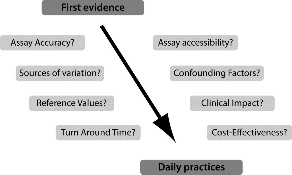
Figure 1. On the way to daily clinical practices: several challenges facing new biomarkers.
First, these biomarkers should first be accessible to the physicians, accurate and allow a short turn around time (TAT) of analysis (70,71). As illustrated in this article, the evidence for emerging biomarkers is often obtained with manual testing. Development of more automated, easily accessible assays is then mandatory for a transfer to routine practices. As example, we can mention the automated testing for BNP and NT-proBNP which can be rapidly performed on large hospital-based platforms as well as on small point-of-care devices (70,72). The galectin-3 assay will be soon available on automated platforms which may facilitate its use. However, the access to the majority of the biomarkers presented in this article remains limited as automated assays and point-of-care testing are not yet available.
Secondly, the assays for these biomarkers should also undergo to a complete analytical evaluation (73). This evaluation will have to determine the most appropriate measurement matrix, the assay imprecision, the limits of blank, the limits of detection and quantitation, as well as the linearity, the potential high-dose hook effect, the storage stability, the cross-reactivity and the potential interferences with and icterus, lipemia and hemolysis. Laboratorians and in vitro diagnostic manufacturers will have also to joint efforts to define appropriate and personalized reference and medical decision limits, to identify the diagnostic time window and the most appropriate clinical application of the biomarkers and their intra- and inter-individual variability (18,23,74).
Third, the question of the potential added value provided by the biomarker will have to be raised. It is clear that natriuretic peptides represent in vitro diagnostic blockbusters, used in daily clinical and laboratory practices, and supporting the diagnosis and prognosis of HF. The biomarker of inflammation and cardiac remodeling may help for the identification of patients at an increased risk of death in the long term. Such biomarkers may also support biomarker-guided treatment (18,19,75). Further evidence is however required before the recommendation of the most appropriate application. However, the level of evidence of Galectin-3, GDF15 and Big ET-1 is rising quickly which may stimulate their prime time use in a near future.
Fourth, the integration of biomarkers from different pathways in multimarker strategies (MMS) should also be considered (76). Thus, Sabatine et al. selected one marker of damage to cardiac tissue, cTnI, one marker of excessive stretching of cardiac tissue, BNP, and one marker of inflammation, CRP and found in a study of 1,635 patients that those with one, two, and three elevated biomarkers had a 2.1-, 3.1-, and 3.7- fold increase in the risk of death, MI or HF by 6 months (77). Similarly, in a study of non-ST elevation acute coronary syndromes (non-ST ACS), Tello-Montoliu et al. studied the levels of TnT, CRP and Nt pro-BNP in 358 patients and found that the MMS strategy identified increased risk of adverse events at 6 months (78). In addition, Zairis et al. investigated the combined prognostic value of admission serum levels of BNP, cTnI and hs-CRP in 577 patients hospitalised because of HF and using a multivariate Cox regression analysis concluded that increasing numbers of elevated biomarkers gradually increased the risk of 31-day cardiac death (79). A same approach integrating plasma biomarkers that reflect determinants of matrix composition identify the presence of left ventricular hypertrophy and diastolic heart failure have also been proposed (80). These are just the early days for MMS and challenges such as the appropriate selection of the biomarkers remain (76).
Finally, the question of cost effectiveness of these emerging biomarkers can not be ignored in the current evaluation process of laboratory tests and remains largely uncovered (81,82). In HF, the economic impact of BNP testing has been evaluated. BNP testing not only resulted in a significant reduction in time to treatment and re-hospitalization rate but also led to substantial costs savings (83,84). For example, Morimoto et al. have conducted a cost-effectiveness analysis using a Markov model of regular BNP measurement in the outpatient setting and showed the QALY (quality adjusted life year) to be longer for the BNP group and the costs of treatment were also lower for the BNP group (85). Such a work remains therefore to be accomplished for the biomarkers presented in this article.
In conclusion, laboratorians and physicians should consider the potential benefits of biomarkers of inflammation and cardiac remodeling for the risk stratification. Nevertheless, multiple challenges including the careful evaluation of analytical and clinical performances as well as their cost-effectiveness should be considered before a translation to daily practices.